
Cervical Radiculopathy Treatment in Burlington: Physiotherapy for Pinched Nerve in Neck Relief
- Folarin Babatunde PT PhD

- Nov 9, 2024
- 4 min read
Updated: Sep 16, 2025
Cogent Rehab Blog
Folarin Babatunde PT PhD MScSEM MScPT BScPT
November 9, 2024

Understanding Cervical Radiculopathy
Cervical radiculopathy, commonly known as a pinched nerve in the neck, occurs when a nerve in the cervical spine becomes compressed or irritated. Cervical radiculopathy is very common in persons 50 to 54 years of age. This condition can cause neck pain, stiffness, radiating arm pain, tingling, weakness, and numbness. Symptoms may worsen with certain neck postures or movements and often interfere with daily activities such as work, driving, or sleeping.
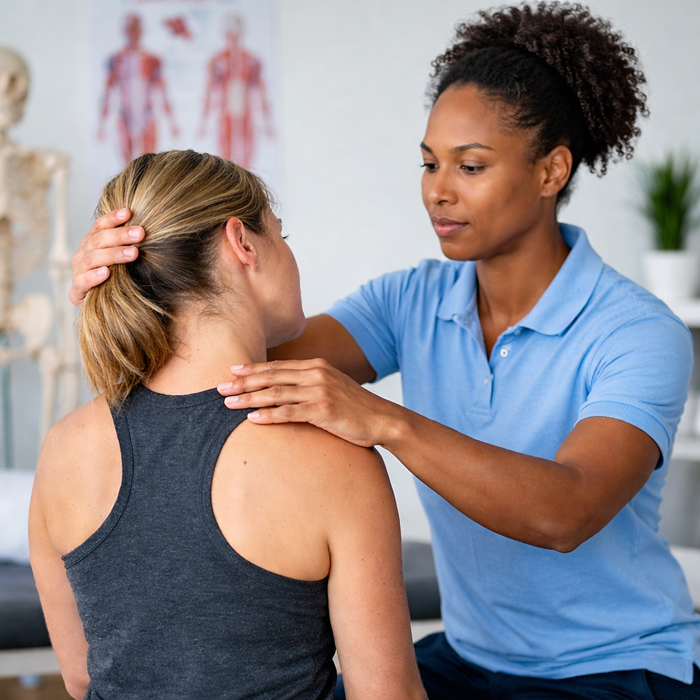
At Cogent Physical Rehabilitation Center in Burlington, our physiotherapists specialize in conservative, evidence-based management of cervical radiculopathy. We provide tailored treatment to relieve pain, restore mobility, and help you return to normal function without surgery.
Don’t let neck and arm pain limit your life — book your assessment at Cogent Rehab Burlington today.
Anatomy of the Cervical Spine

The cervical spine consists of seven vertebrae stacked beneath the skull. Between each vertebra is a disc with two components:
Annulus fibrosus: tough, flexible outer layer
Nucleus pulposus: soft, jelly-like center
These discs act as shock absorbers when you move. Nerve roots branch from the spinal cord through small openings (foramina) in the vertebrae, carrying messages between the brain and body. Radiculopathy occurs when these nerve roots become compressed, often by disc herniation or age-related changes like arthritis.
Who Gets Cervical Radiculopathy?
Cervical radiculopathy is most common in adults aged 40–60, with men slightly more affected than women.
In younger people, it often results from a herniated disc due to lifting, twisting, or sports injuries.
In older adults, it is usually caused by degenerative changes (cervical spondylosis), including disc collapse and bone spur formation.
In rare cases, trauma or posture-related stress may trigger symptoms without a clear injury.
Causes of a Pinched Nerve in the Neck
The most frequent causes include:
Cervical disc herniation – when the soft center of the disc presses against a nerve root
Cervical spondylosis (arthritis) – age-related disc degeneration and bone spur formation narrowing the foramina
Combination of disc herniation and spondylosis – especially in C6 and C7 nerve roots (most common)

Anatomy of pinched nerve in the neck due to disc herniation and cervical spondylosis
Signs and Symptoms of Pinched Nerve in the Neck
Common symptoms include:
Neck pain that radiates into the shoulder or arm
Numbness or tingling in the arm, hand, or fingers
Muscle weakness in the upper limb
Reduced reflexes in the arm
Pain that worsens with certain neck movements
If untreated, chronic nerve irritation can lead to long-term weakness and decreased function.
How Is Cervical Radiculopathy Diagnosed?
Physiotherapists can often diagnose cervical radiculopathy through a detailed history and physical examination.
History: Patients report neck pain radiating to the arm, worsened by neck movements.
Physical exam: Reduced reflexes, muscle weakness, sensory loss, and positive tests (Spurling’s test, arm squeeze test, traction).
Imaging (MRI/CT): Used if symptoms persist or red flags are present (e.g., trauma, malignancy, or infection).

Physiotherapy Treatment for Cervical Radiculopathy
At Cogent Rehab Burlington, our physiotherapists use evidence-based approaches to reduce pain, restore mobility, and improve nerve function.
1. Pain Relief and Inflammation Control
Manual therapy to improve joint mobility
Soft tissue release to reduce muscle tension
Postural correction to decrease nerve irritation
2. Targeted Exercises
Cervical retraction and extension exercises to open nerve pathways
Strengthening exercises for the deep neck flexors and shoulder stabilizers
Nerve gliding techniques to improve neural mobility
3. Education and Ergonomic Advice
Guidance on workplace setup and daily posture
Strategies to avoid aggravating movements
Home exercise programs to support long-term recovery
4. Adjunct Therapies (if appropriate)
Mechanical traction for nerve decompression
Modalities such as IFC (interferential current) for pain relief
Most patients experience improvement without surgery. For cases not responding to conservative care, we work collaboratively with family physicians, neurologists, or orthopedic specialists.
Why Choose Cogent Rehab Burlington for Pinched Nerve in the neck?
Personalized treatment plans tailored to your condition and lifestyle
Evidence-based physiotherapy grounded in current research
Integrated care approach — we collaborate with your healthcare team
Convenient Burlington location with flexible scheduling to reduce wait times
Frequently Asked Questions (FAQ) About Pinched Nerve in the Neck
1. What causes cervical radiculopathy?
Common causes include herniated discs, cervical spondylosis (arthritis of the neck), and bone spurs that narrow the nerve openings.
2. How long does it take to recover?
Many patients improve within 6–12 weeks of physiotherapy, though recovery depends on severity, age, and adherence to treatment.
3. Do I need surgery for a pinched nerve in the neck?
Most cases improve with non-surgical treatments like physiotherapy. Surgery is considered only if symptoms persist or worsen.
4. Can cervical radiculopathy cause permanent damage?
If untreated, chronic compression may lead to long-term weakness or numbness. Early physiotherapy intervention reduces this risk.
Relief is possible without surgery. Start your personalized cervical radiculopathy treatment today at Cogent Rehab Burlington.
Sources
Alshami AM and Bamhair DA. Effect of manual therapy with exercise in patients with chronic cervical radiculopathy: a randomized clinical trial. Tirla.s 2021;22:716.
Borrella-Andrés S. Manual Therapy as a Management of Cervical Radiculopathy: A Systematic Review. Biomed Res Int. 2021;9936981.
Blanpied PR et al. Neck Pain: Revision 2017 Clinical Practice Guidelines Linked to the International Classification of Functioning, Disability and Health From the Orthopaedic Section of the American Physical Therapy Association J Orthop Sports Phys Ther. 2017;47(7):A1-A83
Chiou-Tann FY. Musculoskeletal mimics of cervical radiculopathy. Muscle Nerve. 2022;66:6-14.
Eubanks JD. Cervical Radiculopathy: Nonoperative Management of Neck Pain and Radicular Symptoms. Am Fam Physician. 2010;81:33-40
Rafiq S. et al. Comparison of neural mobilization and conservative treatment on pain, range of motion, and disability in cervical radiculopathy: A randomized controlled trial. PLoS One 2022;17:e0278177.
Sleijser‑Koehorst MLS. et al. Clinical course and prognostic models for the conservative
management of cervical radiculopathy: a prospective cohort study. Eur Spine J. 2018;27:2710–2719
Figure 3: https://my.clevelandclinic.org/health/diseases/17685-cervical-spondylosis


Comments